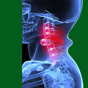
Vertebral migration is an alternative diagnostic terminology for spondylolisthesis. However, the concept is complicated to understand and some diagnosed patients obviously do not have a true comprehension of the nature of the condition or its potential effects. In order to continue our patient education efforts, we have decided to create this article to help people to better visualize what spondylolisthesis is all about from a completely different point of view.
This discussion provides a simple and straight-forward explanation of listhesis conditions in the human spine, including descriptions that can help every patent to better realize the causes and consequences of vertebral listhesis. We will also use the opportunity to present a fact-based analysis of the degrees of listhesis in order to help patients to enjoy a more accurate prognosis to the future state of their spinal condition.
Vertebral Migration and Spinal Curvature
We have already described and defined spondylolisthesis from a scientific point of view elsewhere in this website. However, not all patients fully understand the diagnosis and so we will redefine it here for the sake of simplicity:
The human spine contains 24 individual vertebrae (not including the various fused bones of the sacrum and coccyx). These are the bones which are susceptible to spondylolisthesis development. The normal human spine should be straight when viewed from the front or back, but will demonstrate a variety of curvatures when viewed from the side. These curvatures begin in the neck with what is called lordosis. Cervical lordosis describes a gentle curvature in the neck wherein the open side of the curvature faces the rear of the body. As the spine transitions to the upper back, the curvature reverses direction, becoming thoracic kyphosis. This kyphosis describes a gentle curvature of the upper and middle back areas with the open side of the curvature facing the front of the body. As we transition again to the lower back, the curvature changes once again to lordosis. This lumbar lordosis once again features the open end of the curvature facing the rear of the body.
As the spine progresses from the neck to the tailbone, the individual vertebral bones each move a bit from front to back, as well as tilting slightly to facilitate these curvatures. Picture a gentle wave effect of bones cascading down in 3 distinct curvature patterns and you can accurately visualize the cervical, thoracic and lumbar spinal regions, also called the neck, upper-middle back and lower back. It is important to understand these curvatures in order to truly understand spondylolisthesis.
Spinal Bone Migration
Now you can imagine the spinal column as an arrangement of curvatures composed of separate, but related, vertebral bones. In a normal spine, each bone will have a distinct location where it will reside in relation to the bone above and below it. This location is determined by the position where the bone is located in the curvature. Some vertebral bones will be on the ascending part of the curvature, while others will be on the descending aspect of the curvature. A few bones will fall in the apex of the curvature, where it reaches its maximum degree of lordosis or kyphosis, while others will be at the frontiers where the curvature transitions from lordosis to kyphosis and vice versa. While all of this sounds rather complicated, it is all quite simple. Just look at a picture of the spine and it all becomes crystal clear.
The relative position of each vertebral bone in relation to its neighbors is predictable and well defined in a normal spine. The bones will be positioned following the curves and will each be spaced typically from each other in terms of front to back proximity.
Spondylolisthesis occurs when one or more of these vertebral bones “slips” out of place in the spine and falls out of alignment with the rest of the bones above and/or below. The vertebral bone might move excessively forward of its neighbors in a condition called anterolisthesis. The bone might also move excessively backwards in compared to its neighbors, in a condition called retrolisthesis. If no direction is specified, most cases of spondylolisthesis should be considered to be anterolisthesis, as this is a much more common profile. So in essence, spondylolisthesis consists of abnormal vertebral positioning out of alignment with the expected curvatures of the spine.
Vertebral Migration Factsheet
All of this can sound really, really bad to laymen, since having a spinal bone out of place sounds like the perfect cause of back pain and potentially worse problems. However, this assumption does not necessarily represent the facts of listhesis.
Spondylolisthesis is graded into 4 categories according to severity. Lower grades of 1 and 2 represent minor and moderate degrees of abnormal positioning in relation to the expected normal placement of the spinal bone. Grades 3 and 4 represent severe and extreme misalignments of the vertebral bone in relation to the surrounding vertebrae. This diagnosis of classification can be made using any type of medical imaging, including x-ray, MRI or CT scan, but is usually easiest to visualize using basic x-ray films from the sagittal (also called lateral) view. Basically this means the images are taken from the side of the body.
You can learn more about the various spondylolisthesis gradings and the expected consequences of each by reading our dedicated coverage of each topic. We hope that this essay paints a more vivid and understandable picture of listhesis in a way that makes sense to you. Knowledge is the best weapon against back or neck pain. Understanding your diagnosis is step one in the ongoing process of personal enrichment.
Spondylolisthesis > Isthmic Spondylolisthesis > Vertebral Migration


